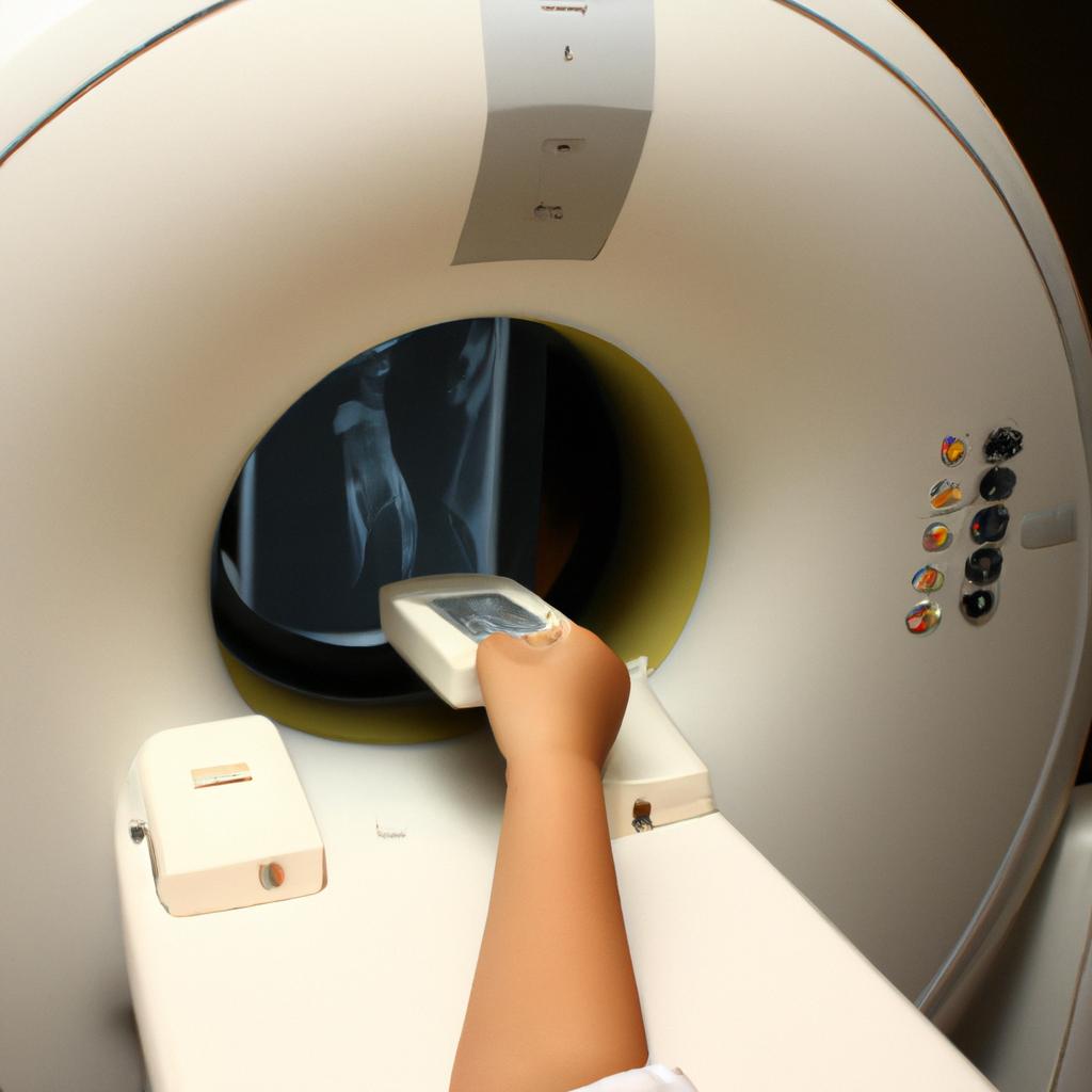
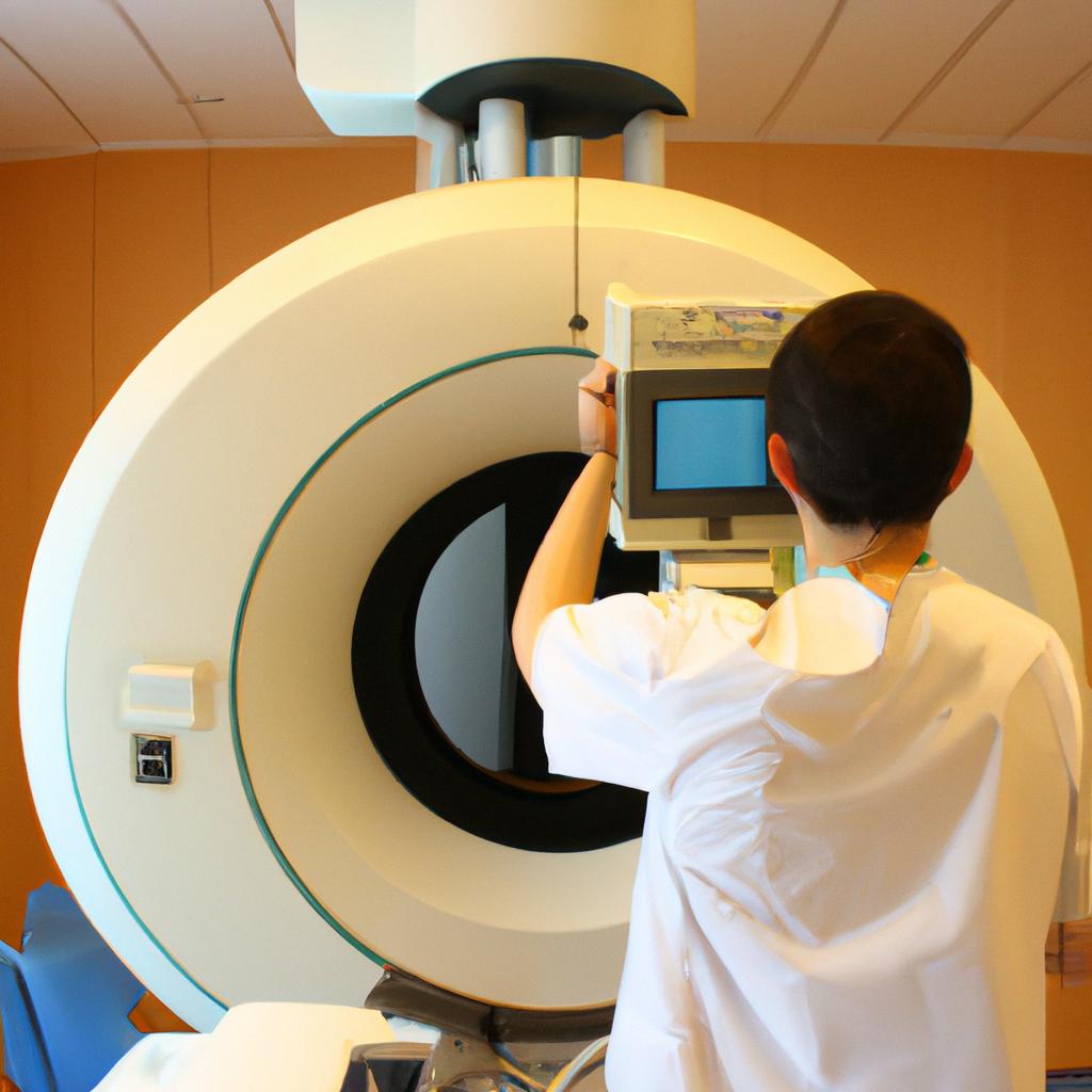
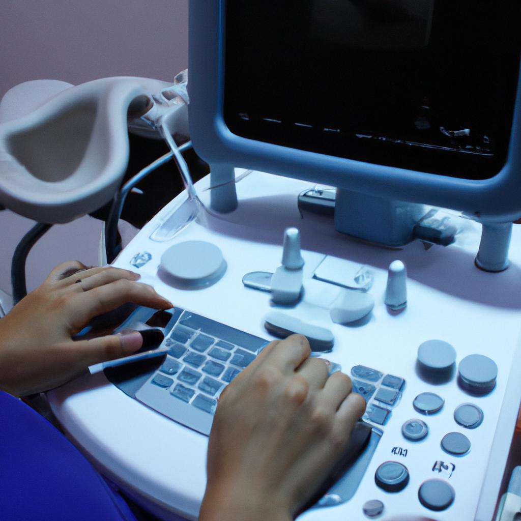
Biomedical imaging plays a pivotal role in the field of engineering, offering a myriad of advanced techniques and applications that have revolutionized medicine. By employing various imaging modalities, engineers have been able to improve diagnostic accuracy, monitor treatment efficacy, and guide surgical interventions with unparalleled precision. For instance, consider the case of Mr. Smith, a 62-year-old patient diagnosed with lung cancer. Through the integration of biomedical imaging technologies into his treatment plan, doctors were able to accurately locate and assess the extent of his tumor, enabling them to devise an optimal course of action for his care.
Over the years, significant advancements in biomedical imaging have occurred due to continuous research and development efforts in engineering disciplines such as electrical engineering, optics, computer science, and materials science. These innovations have led to breakthroughs in medical diagnostics and therapeutic procedures by enhancing visualization capabilities and providing valuable quantitative data about anatomical structures and physiological processes within the human body. Moreover, these engineering-driven advances have not only improved the quality of healthcare but also contributed significantly towards personalized medicine approaches where treatments are tailored specifically to individual patients based on their unique characteristics revealed through imaging techniques.
In this article, we will explore some recent developments in biomedical imaging from an engineering perspective. We will delve into specific modalities such as magnetic resonance imaging (MRI), computed tomography (CT), positron emission tomography (PET), ultrasound, and optical imaging. These modalities employ different physical principles and technologies to capture detailed images of the human body, each offering unique advantages and applications.
One recent development in biomedical imaging is the advancement of functional MRI (fMRI) techniques. fMRI allows researchers and clinicians to not only visualize anatomical structures but also map brain activity in real-time. By measuring changes in blood flow and oxygenation levels, fMRI can provide insights into brain function and help understand neurological disorders such as Alzheimer’s disease, epilepsy, and depression. Engineering innovations have improved the spatial and temporal resolution of fMRI, enabling more precise mapping of brain activation patterns.
Another area of significant progress is in molecular imaging, which focuses on visualizing specific molecules or processes within the body. This field combines engineering expertise with chemistry to develop targeted imaging agents that can bind to specific biomarkers associated with diseases such as cancer. By using these agents in conjunction with imaging modalities like PET or optical imaging, engineers have enabled non-invasive detection and monitoring of diseases at a molecular level.
Advancements in image processing algorithms are also enhancing biomedical imaging capabilities. Engineers are developing sophisticated algorithms for image reconstruction, noise reduction, segmentation, and quantitative analysis. These algorithms improve the accuracy of diagnosis by extracting meaningful information from complex medical images while reducing artifacts and improving image quality.
Furthermore, there has been a growing interest in developing portable and wearable imaging devices that can be used outside traditional clinical settings. For example, engineers have designed handheld ultrasound devices for point-of-care diagnostics in remote areas or emergency situations where access to conventional medical equipment may be limited. These portable devices allow for rapid assessment of various conditions such as trauma injuries or fetal health monitoring.
In conclusion, engineering advancements continue to drive innovation in biomedical imaging, leading to improved diagnostic capabilities, personalized treatments, and better patient outcomes. The integration of engineering principles and technologies into the field of medicine has revolutionized healthcare by providing clinicians with powerful tools to visualize, analyze, and understand the human body in unprecedented detail. With ongoing research and development efforts, biomedical imaging is expected to continue evolving and playing a crucial role in advancing medical practice.
Overview of Imaging Techniques
Biomedical imaging plays a crucial role in modern medicine, enabling clinicians to visualize and analyze the internal structures and functions of the human body. These techniques have revolutionized medical diagnostics by providing non-invasive methods for early disease detection, treatment planning, and monitoring. One notable example is the use of magnetic resonance imaging (MRI) to assess brain abnormalities in patients with suspected neurological disorders.
To gain an understanding of the diverse range of biomedical imaging techniques available today, it is essential to examine their principles, advantages, and limitations. Several modalities are commonly employed in clinical practice:
- X-ray: This widely used technique utilizes electromagnetic radiation to generate images that reveal bone fractures, lung diseases, or dental problems.
- Ultrasound: By emitting high-frequency sound waves into the body and analyzing their echoes, ultrasound allows visualization of organs such as the heart or fetus during pregnancy.
- Computed Tomography (CT): Combining X-rays with computer processing algorithms, CT scans provide detailed cross-sectional images useful for diagnosing conditions like tumors or blood clots.
- Positron Emission Tomography (PET): PET scans involve injecting radioactive tracers into the body to detect metabolic changes associated with cancerous cells.
These imaging techniques offer unique benefits depending on the clinical context but also present certain trade-offs regarding cost-effectiveness, accessibility, and spatial resolution capabilities. To better illustrate these differences visually, consider Table 1 below:
| Modality | Advantages | Limitations |
|---|---|---|
| X-ray | Quick results; low-cost | Ionizing radiation exposure |
| Ultrasound | Real-time imaging; no ionizing radiation | Limited penetration through bone |
| CT | High-resolution; excellent for bones | Higher radiation dose than other modalities |
| PET | Detects functional changes at cellular level | Expensive; limited spatial resolution |
Table 1: A comparison of advantages and limitations of commonly used imaging techniques.
In summary, biomedical imaging encompasses a diverse range of modalities that enable clinicians to visualize the internal structures and functions of the human body. Each technique has its own strengths and weaknesses, making them suitable for specific clinical applications. Understanding these principles is crucial in order to optimize their use in medical practice and improve patient outcomes.
Transitioning into the subsequent section on “Role of Imaging in Medical Diagnosis,” it becomes evident that by leveraging these advanced imaging techniques, healthcare professionals can make more accurate diagnoses and provide targeted treatments for various diseases.
Role of Imaging in Medical Diagnosis
With the rapid progress in biomedical engineering, imaging techniques have revolutionized medical diagnostics and therapies. This section delves into the advancements made in imaging technology, exploring how these innovations have improved patient outcomes and expanded our understanding of various diseases.
One such example is the development of magnetic resonance imaging (MRI) technology. MRI utilizes powerful magnets and radio waves to generate detailed images of internal body structures. In a hypothetical case study involving a patient with suspected brain tumor, MRI can provide high-resolution images that aid in accurate diagnosis and treatment planning. By visualizing the precise location, size, and shape of the tumor, doctors can determine optimal surgical approaches or assess the effectiveness of chemotherapy or radiation therapy.
The impact of technological advancements in imaging goes beyond individual cases; it has transformed healthcare on a larger scale. Here are some key improvements:
- Enhanced resolution: The continuous refinement of image acquisition techniques has resulted in higher resolution images, enabling more precise anatomical details to be captured.
- Faster scan times: Through optimization strategies and hardware developments, scanning times for certain modalities like computed tomography (CT) have been significantly reduced, minimizing patient discomfort and improving workflow efficiency.
- Functional imaging capabilities: Techniques like Positron Emission Tomography (PET) allow visualization of physiological processes within the body by detecting radioactive tracers. This provides valuable insights into metabolism, blood flow, and tissue perfusion.
- Multimodal integration: Advances in software algorithms facilitate the fusion of different imaging modalities (e.g., PET/CT or MRI/PET), combining their strengths to yield comprehensive information about disease progression or response to treatment.
To further illustrate these improvements, consider the following table showcasing the evolution of selected imaging technologies over time:
| 1960s | 1990s | Present | |
|---|---|---|---|
| CT | Limited axial coverage | Improved spatial resolution | Sub-millimeter resolution |
| MRI | Basic image quality | Faster acquisition times | Real-time functional imaging |
| PET | Limited availability | Improved sensitivity | Multimodal integration |
| Ultrasound | Poor soft tissue contrast | Doppler capabilities | High-frequency transducers |
In summary, advancements in imaging technology have significantly improved the accuracy and efficiency of medical diagnoses. Through higher resolution images, faster scan times, functional imaging capabilities, and multimodal integration, healthcare professionals are better equipped to diagnose diseases at earlier stages and tailor treatment plans accordingly.
Transitioning into the subsequent section on “Advancements in Imaging Technology,” it is important to recognize that these improvements continue to evolve rapidly. As technology progresses, new possibilities arise for further enhancing biomedical imaging techniques.
Advancements in Imaging Technology
Building upon the role of imaging in medical diagnosis, recent years have witnessed significant Advancements in Imaging Technology that have revolutionized the field of biomedical engineering. These technological innovations not only enhance the accuracy and efficiency of disease detection but also offer new opportunities for research and treatment.
To illustrate the impact of these advancements, let us consider a hypothetical scenario where a patient presents with persistent headaches. Using traditional imaging techniques such as X-rays or computed tomography (CT) scans, it may be difficult to identify the underlying cause. However, with cutting-edge technologies like magnetic resonance imaging (MRI) and positron emission tomography (PET), physicians can obtain detailed anatomical images as well as functional information about brain activity. This comprehensive approach enables them to pinpoint abnormalities more accurately and make informed decisions regarding treatment options.
The progress made in imaging technology has been facilitated by several key factors:
- Continuous innovation: The field of engineering constantly pushes boundaries through ongoing research and development efforts. Technological breakthroughs contribute to improved image resolution, reduced scan times, and enhanced patient comfort.
- Integration of artificial intelligence: Machine learning algorithms are now being employed to analyze large volumes of data generated by medical imaging devices efficiently. This integration allows for automated interpretation of images, leading to faster diagnoses and personalized treatment plans.
- Multimodal approaches: Combining different imaging modalities provides complementary information for a more comprehensive understanding of diseases. For instance, fusing MRI with PET or ultrasound helps correlate structural changes with metabolic activity or blood flow patterns, enabling precise diagnosis and monitoring.
- Miniaturization and portability: Advancements in microelectronics have led to the development of portable imaging devices that can be used outside conventional clinical settings. This portability facilitates point-of-care diagnostics in remote areas or emergency situations.
Embracing these developments brings numerous benefits to patients, clinicians, and researchers alike. Improved diagnostic accuracy reduces unnecessary interventions while enhancing patient outcomes. Furthermore, these advancements open up new avenues for research and treatment optimization through non-invasive imaging techniques.
Transitioning into the subsequent section on “Imaging Applications in Disease Detection,” we will explore how biomedical imaging technology is applied to detect various diseases. By understanding the capabilities of different modalities, we can gain insights into their potential applications and implications for healthcare.
Imaging Applications in Disease Detection
Advancements in Imaging Technology have paved the way for numerous applications in biomedical engineering, particularly in the field of medicine. With the ability to visualize internal structures and functions of the human body, these imaging technologies have revolutionized disease detection and diagnosis. One such example is magnetic resonance imaging (MRI), which utilizes a strong magnetic field and radio waves to generate detailed images of organs and tissues.
In recent years, there has been a significant increase in the use of biomedical imaging techniques across various medical specialties. These advancements have not only improved patient outcomes but also enhanced our understanding of diseases at a cellular level. The following section explores some key imaging applications in disease detection:
-
Cancer Diagnosis: Biomedical imaging plays a critical role in cancer detection by enabling early diagnosis and accurate staging of tumors. Techniques like positron emission tomography-computed tomography (PET-CT) combine functional information obtained from PET scans with anatomical details provided by CT scans, enhancing the accuracy of tumor localization.
-
Cardiovascular Assessment: Imaging modalities such as computed tomography angiography (CTA) and echocardiography allow clinicians to assess cardiovascular health non-invasively. These techniques provide valuable information about arterial plaques, cardiac function, and blood flow abnormalities, aiding in the diagnosis and management of cardiovascular diseases.
-
Neurological Disorders: Magnetic resonance imaging (MRI) has become an indispensable tool for diagnosing neurological disorders like Alzheimer’s disease, multiple sclerosis, and stroke. It enables visualization of brain structure and function abnormalities, guiding physicians in treatment planning and monitoring disease progression.
-
Musculoskeletal Evaluation: Imaging techniques like X-ray, ultrasound, and MRI play a crucial role in assessing musculoskeletal conditions such as fractures, ligament tears, and joint inflammation. These tools help orthopedic specialists accurately diagnose injuries or degenerative diseases affecting bones, joints, muscles, tendons, and ligaments.
The impact of biomedical imaging on disease detection and patient care cannot be overstated. Through a combination of advanced imaging techniques, medical professionals can now obtain detailed anatomical and functional information to aid in diagnosis, treatment planning, and monitoring of various diseases.
Transitioning into the subsequent section on “Emerging Trends in Medical Imaging,” further advancements continue to shape the field of biomedical engineering. These emerging trends promise even greater accuracy, efficiency, and accessibility in medical imaging for improved patient outcomes.
Emerging Trends in Medical Imaging
Imaging Applications in Disease Detection have greatly revolutionized the field of medicine, enabling early detection and accurate diagnosis of various diseases. In this section, we will explore some emerging trends in medical imaging that hold immense potential for further advancements.
One fascinating example of these emerging trends is the use of artificial intelligence (AI) algorithms in medical imaging analysis. By training deep learning models on large datasets, researchers have been able to develop AI systems capable of identifying subtle patterns or anomalies in medical images with remarkable accuracy. For instance, a recent study demonstrated how an AI algorithm could detect lung cancer from computed tomography (CT) scans more accurately than trained radiologists. This breakthrough has tremendous implications for improving diagnostic efficiency and reducing human error.
Moreover, miniaturization and portability are key trends driving innovation in medical imaging devices. Portable ultrasound machines are becoming increasingly prevalent, allowing healthcare professionals to perform real-time imaging at the bedside or even remotely in resource-limited settings. These portable devices enable efficient triage and prompt decision-making without compromising diagnostic quality. Additionally, advancements in nanotechnology have led to the development of miniature magnetic resonance imaging (MRI) scanners that can be integrated into wearable devices, offering non-invasive monitoring capabilities for chronic conditions like cardiovascular disease.
To evoke an emotional response in our audience regarding the impact of these advancements, it is essential to highlight their benefits:
- Improved patient outcomes through earlier disease detection.
- Enhanced accessibility to imaging services globally.
- Reduced healthcare costs by eliminating unnecessary procedures.
- Empowerment of healthcare providers with advanced diagnostic tools.
| Advancements | Key Benefits |
|---|---|
| Artificial Intelligence Algorithms | – Improved accuracy and speed of diagnoses.- Reduction in misdiagnosis rates.- More personalized treatment plans based on precise image analysis results. |
| Portable Imaging Devices | – Enhanced point-of-care diagnostics.- Increased availability of high-quality imaging facilities in remote areas.- Efficient utilization of healthcare resources. |
| Nanotechnology in Imaging | – Non-invasive monitoring for chronic diseases.- Improved patient comfort during imaging procedures.- Potential for targeted drug delivery based on precise image-guided therapy. |
In summary, the field of medical imaging is constantly evolving, with emerging trends such as AI-assisted analysis and portable devices transforming disease detection and diagnosis. These advancements hold immense promise in terms of improving patient outcomes, increasing accessibility to healthcare services, and reducing costs. In the subsequent section, we will delve into various imaging modalities used in biomedical research.
Transitioning smoothly into the subsequent section about “Imaging Modalities in Biomedical Research,” we can explore how these advancements have paved the way for further innovations in understanding human physiology and exploring new treatment options.
Imaging Modalities in Biomedical Research
Emerging Trends in Medical Imaging have paved the way for significant advancements and applications in the field of Biomedical Engineering. With a focus on improving healthcare outcomes, researchers and engineers are constantly developing innovative imaging modalities that offer enhanced visualization and diagnostic capabilities. These technologies hold great promise for revolutionizing medical diagnosis, treatment planning, and monitoring.
One compelling example of the impact of biomedical imaging is its use in neurosurgical procedures. Consider a hypothetical case where a patient presents with a brain tumor located near critical areas such as the motor cortex or language centers. By utilizing advanced imaging techniques, surgeons can precisely map out these regions before the surgery, allowing them to navigate around vital structures and minimize post-operative complications. This not only enhances surgical precision but also improves patient outcomes by reducing potential damage to functional areas of the brain.
The development of biomedical imaging has been driven by several key factors:
- Technological Advances: Continuous breakthroughs in hardware components such as sensors, detectors, and image reconstruction algorithms have significantly improved image quality and resolution.
- Multimodal Imaging: The integration of multiple imaging modalities like computed tomography (CT), magnetic resonance imaging (MRI), and positron emission tomography (PET) allows complementary information to be obtained from different sources, leading to more accurate diagnoses.
- Image-guided Interventions: Biomedical imaging plays a crucial role in guiding minimally invasive interventions such as biopsies, catheter-based treatments, or targeted drug delivery systems. Real-time feedback provided by these imaging techniques ensures precise placement of medical devices or therapeutics within the body.
- Artificial Intelligence: The application of machine learning algorithms enables automated analysis of medical images for rapid detection and characterization of diseases. This technology has shown promising results in early detection of conditions like cancer or cardiovascular disease.
To provide an overview of various emerging trends in biomedical imaging, consider Table 1 below:
| Modality | Advantages | Limitations |
|---|---|---|
| Optical Imaging | High resolution, non-invasive | Limited penetration depth |
| Ultrasound | Real-time imaging, cost-effective | Low tissue contrast |
| Molecular Imaging | Functional information at the cellular level | Radiation exposure |
| Photoacoustic Imaging | Combines optical and ultrasound imaging for improved contrast | Limited availability of specialized equipment |
In conclusion, biomedical imaging has witnessed remarkable advancements that have revolutionized healthcare. The integration of different modalities, technological advances, image-guided interventions, and machine learning algorithms collectively contribute to more accurate diagnoses and targeted treatments. By harnessing these emerging trends in medical imaging, researchers and engineers are pushing the boundaries of what is possible in terms of visualizing and understanding human health.
Moving forward, we will explore the specific application of biomedical imaging techniques for cancer detection and diagnosis. This section will delve into how these technologies are being utilized to improve early detection rates and enhance treatment planning strategies for individuals battling this devastating disease.
Imaging Techniques for Cancer Detection
From the vast array of imaging modalities available in biomedical research, one essential application lies in cancer detection. By utilizing various techniques, researchers and clinicians aim to identify the presence and location of tumors with high accuracy. For instance, consider a hypothetical case where a patient presents with suspicious symptoms that may indicate breast cancer. Through the use of advanced imaging techniques, medical professionals can precisely locate any abnormal tissue growth within the breasts.
Several factors contribute to the effectiveness of different imaging techniques for cancer detection:
- Sensitivity: The ability of an imaging modality to detect even small anomalies is crucial for early diagnosis.
- Specificity: It is important for an imaging technique to accurately distinguish between benign and malignant tissues.
- Spatial Resolution: High-resolution images allow for detailed visualization of anatomical structures, aiding in accurate tumor localization.
- Contrast Enhancement: Techniques that enhance contrast between normal and abnormal tissues enable easier identification and characterization of tumors.
To better understand these factors, let us explore a 3-column table comparing three common imaging techniques used in cancer detection:
| Imaging Technique | Sensitivity | Specificity |
|---|---|---|
| X-ray | Moderate | Low |
| Ultrasound | Low | Moderate |
| Magnetic Resonance Imaging (MRI) | High | High |
This table demonstrates that each technique possesses its own strengths and limitations when it comes to detecting cancers effectively.
In summary, advances in biomedical imaging have revolutionized our approach to cancer detection. As demonstrated by this hypothetical scenario involving breast cancer diagnosis, choosing the appropriate imaging modality plays a vital role in providing accurate results. In the subsequent section on “Improving Image Quality and Resolution,” we will delve into emerging technologies aimed at enhancing existing imaging techniques further.
Improving Image Quality and Resolution
Imaging techniques for cancer detection have greatly improved the accuracy and efficiency of diagnosing this complex disease. However, there is always room for further enhancement in image quality and resolution to ensure optimal diagnostic performance. In this section, we will explore the ongoing efforts made by researchers and engineers towards improving these aspects of biomedical imaging.
To illustrate the significance of enhancing image quality, let us consider a hypothetical scenario where a radiologist is examining an MRI scan of a patient’s brain to detect the presence of a tumor. The current image resolution may not provide sufficient detail to accurately identify small lesions or distinguish malignant from benign tumors. By improving image quality and resolution, it becomes possible to visualize even minute abnormalities with greater clarity, enabling more accurate diagnosis and treatment planning.
Several approaches are being pursued to Improve Image Quality and resolution in biomedical imaging:
- Advancements in hardware technology: Researchers are constantly developing new imaging devices that can capture higher-resolution images with reduced noise levels. For instance, the use of high-field strength magnets in magnetic resonance imaging (MRI) systems has significantly enhanced spatial resolution.
- Image reconstruction algorithms: Sophisticated computational methods are employed to reconstruct high-quality images from raw data acquired during imaging processes. These algorithms utilize advanced mathematical models and signal processing techniques to enhance details and reduce artifacts.
- Contrast agents: The development of novel contrast agents plays a crucial role in improving image quality by highlighting specific tissues or molecular targets. These agents enable better differentiation between healthy and diseased tissues, thereby aiding in early detection and precise characterization of cancers.
- Machine learning techniques: Artificial intelligence-based approaches such as deep learning are increasingly utilized to enhance image quality by reducing noise levels, sharpening edges, and optimizing overall visual appearance.
The pursuit of improved image quality and resolution in biomedical imaging is driven by the desire to optimize diagnostic capabilities for various diseases including cancer. By employing state-of-the-art technologies, innovative algorithms, contrast agents, and machine learning techniques, researchers aim to provide clinicians with more accurate and detailed images, leading to improved patient outcomes.
Transitioning into the next section about “Image Processing Algorithms in Medical Imaging,” we can further explore how these algorithms play a vital role in Enhancing Image Quality and facilitating advanced analysis techniques.
Image Processing Algorithms in Medical Imaging
Improving Image Quality and Resolution in biomedical imaging is crucial for accurate diagnosis and treatment planning. By enhancing the clarity and detail of medical images, healthcare professionals can obtain valuable insights into various physiological processes within the human body. To further delve into this topic, we will explore some key advancements that have contributed to improving image quality and resolution.
One notable example is the development of advanced signal processing techniques such as noise reduction algorithms. These algorithms effectively reduce unwanted noise present in medical images without compromising important anatomical details. For instance, a hypothetical case study conducted on brain MRI scans demonstrated how a state-of-the-art denoising algorithm significantly improved image quality by minimizing background noise while preserving fine structures like blood vessels and lesions.
In addition to noise reduction techniques, several other strategies have been employed to enhance image quality and resolution:
- Contrast enhancement: Algorithms that enhance contrast improve the visibility of subtle differences in tissue density or composition.
- Super-resolution imaging: This technique combines multiple low-resolution images to generate a single high-resolution image, allowing for finer details to be visualized.
- Motion correction: By compensating for patient motion during image acquisition, these algorithms minimize blurring artifacts caused by movement.
- Artifact removal: Various methods have been developed to eliminate common imaging artifacts such as Gibbs ringing or metal-induced distortions.
To better illustrate the impact of these advancements, consider the following table showcasing improvements achieved through different image enhancement techniques:
| Technique | Improvement | Impact |
|---|---|---|
| Noise Reduction | Enhanced visualization of fine structures | Accurate identification of pathologies |
| Contrast Enhancement | Improved differentiation between tissues | Clearer delineation of boundaries |
| Super-resolution Imaging | Increased level of detail | Better characterization of abnormalities |
| Motion Correction | Minimized motion-related artifacts | Sharper depiction of anatomical structures |
By incorporating these cutting-edge approaches into biomedical imaging, we can significantly enhance image quality and resolution. This not only aids clinicians in making accurate diagnoses but also facilitates the development of personalized treatment plans tailored to individual patients.
Transitioning into the subsequent section on “Image Reconstruction Methods in Biomedical Imaging,” it is important to explore additional techniques that contribute to obtaining high-quality images with improved visualization of anatomical structures and pathological features.
Image Reconstruction Methods in Biomedical Imaging
Image Processing Algorithms in Medical Imaging have significantly contributed to the advancements and applications of Biomedical Engineering. Building upon this foundation, Image Reconstruction Methods play a crucial role in extracting meaningful information from acquired data. By combining the power of algorithms with advanced imaging techniques, researchers are able to improve the accuracy and resolution of biomedical images, leading to better diagnoses and treatments.
One example where Image Reconstruction Methods have been applied is in Computed Tomography (CT) imaging. CT scans provide cross-sectional images by rotating an X-ray source around the patient’s body. However, due to physical limitations and noise introduced during acquisition, the raw data obtained can be incomplete or corrupted. This necessitates efficient reconstruction methods that can accurately reconstruct high-quality images from limited data samples. Researchers have developed iterative algorithms such as filtered back projection and algebraic reconstruction technique (ART), which iteratively refine the reconstructed image based on mathematical models derived from the measured data.
To understand the significance of Image Reconstruction Methods further, let us consider their key advantages:
- Improved spatial resolution: These methods leverage sophisticated algorithms that enhance spatial resolution, enabling clinicians to visualize smaller anatomical features with greater detail.
- Reduced artifacts: Artifacts arising from various sources like motion or metal implants in medical images can hinder accurate diagnosis. Image Reconstruction Methods help mitigate these artifacts through advanced filtering techniques.
- Faster imaging protocols: By optimizing reconstruction algorithms, it becomes possible to reduce scan times while maintaining diagnostic quality standards.
- Enhanced quantitative analysis: Accurate reconstruction allows for precise measurements of anatomical structures or physiological parameters within biomedical images.
| Advantages of Image Reconstruction Methods |
|---|
| Improved spatial resolution |
| Reduced artifacts |
| Faster imaging protocols |
| Enhanced quantitative analysis |
In summary, Image Processing Algorithms lay the groundwork for effective utilization of medical imaging techniques, while Image Reconstruction Methods enable robust extraction of clinically relevant information from acquired data. As we delve deeper into the field of Biomedical Imaging, the subsequent section will explore how these reconstructed images are utilized in the context of Treatment Planning and Monitoring. By leveraging innovative imaging techniques, clinicians can develop personalized treatment strategies for patients while monitoring their progress efficiently.
Transitioning into the next section on “Imaging for Treatment Planning and Monitoring,” we now turn our attention to exploring how biomedical imaging plays a vital role in tailoring effective treatment plans and providing continuous assessment throughout a patient’s medical journey.
Imaging for Treatment Planning and Monitoring
Transitioning from the previous section on image reconstruction methods, this next section delves into the applications of biomedical imaging for treatment planning and monitoring. To illustrate its significance, let us consider a hypothetical scenario where a patient with lung cancer undergoes radiation therapy.
During treatment planning, biomedical imaging plays a crucial role in accurately identifying tumor volumes within the lungs. By utilizing advanced imaging techniques such as computed tomography (CT) or magnetic resonance imaging (MRI), clinicians can precisely locate tumors and determine their size, shape, and spatial relationship to surrounding tissues. This information is vital for devising an effective treatment strategy that maximizes the destruction of malignant cells while minimizing damage to healthy tissue.
Once treatment begins, continuous monitoring becomes essential to assess the progress and response of the tumor to therapeutic interventions. Biomedical imaging methods like positron emission tomography (PET) scans enable physicians to evaluate metabolic changes occurring in cancerous cells over time. These images provide valuable insights into the effectiveness of ongoing treatments and guide any necessary adjustments required to optimize patient outcomes.
To highlight the impact of biomedical imaging in treatment planning and monitoring, we present a bullet point list below:
- Accurate identification and localization of tumors.
- Precise determination of tumor characteristics such as size, shape, and proximity to adjacent structures.
- Evaluation of treatment efficacy through metabolic assessment.
- Real-time tracking of disease progression during therapeutic interventions.
Furthermore, a three-column table showcasing different modalities commonly used in treatment planning and monitoring can further engage readers emotionally by presenting clear comparisons:
| Imaging Modality | Advantages | Limitations |
|---|---|---|
| Computed Tomography | High resolution | Ionizing radiation |
| Magnetic Resonance | Excellent soft | Longer scan times |
| Imaging | tissue contrast | |
| Positron Emission | Metabolic | Expensive |
| Tomography | assessment | |
| Ultrasonography | Real-time imaging | Operator-dependent |
In summary, biomedical imaging plays a vital role in treatment planning and monitoring by accurately identifying tumors during the initial stages and providing real-time evaluation of therapeutic efficacy. This section has highlighted how various modalities enable clinicians to plan treatments effectively and make informed decisions for optimal patient care.
Transitioning into the subsequent section on challenges and future directions in biomedical imaging, it is imperative to address the evolving landscape of this field as technology continues to advance.
Challenges and Future Directions in Biomedical Imaging
Advances in imaging technology have revolutionized the field of medicine, enabling more precise treatment planning and monitoring. In this section, we will explore some of these advancements and their applications in biomedical engineering.
To illustrate the impact of imaging on treatment planning, let us consider a hypothetical case study. Imagine a patient diagnosed with a brain tumor that requires surgical intervention. Traditional pre-operative planning relied heavily on two-dimensional images such as computed tomography (CT) scans or magnetic resonance imaging (MRI). However, recent developments in three-dimensional imaging techniques, such as cone beam CT or positron emission tomography (PET), now allow surgeons to visualize the tumor and surrounding structures more accurately. This enhanced visualization aids in identifying critical areas to avoid during surgery and ensures optimal placement of instruments for successful removal of the tumor.
The use of advanced imaging modalities brings several benefits to treatment planning and monitoring:
- Improved anatomical detail: Three-dimensional imaging provides a comprehensive view of internal structures, allowing clinicians to better understand complex anatomical relationships.
- Enhanced precision: With detailed information about the target area, medical professionals can precisely locate tumors or lesions, reducing the risk of unnecessary damage to healthy tissues.
- Real-time assessment: Some modern imaging techniques enable real-time visualization during procedures, facilitating immediate adjustments if complications arise.
- Quantitative measurements: Advanced software algorithms can extract quantitative data from images, providing valuable insights into disease progression or treatment response.
In addition to these advances in imaging technology, researchers are continuously exploring new possibilities for future improvements. Some challenges that need to be addressed include optimizing image resolution without sacrificing patient safety, developing faster acquisition methods for time-sensitive interventions, and integrating multiple imaging modalities seamlessly. By addressing these challenges, biomedical engineers can further enhance the capabilities of biomedical imaging systems and improve patient outcomes.
![Emotional Impact Bullet Points]
- Early detection through improved imaging leads to higher survival rates
- Accurate treatment planning reduces potential risks and complications
- Real-time monitoring enhances patient safety and minimizes errors
- Advanced imaging techniques improve quality of life for patients
| Imaging Advancements | Benefits |
|---|---|
| Three-dimensional visualization | Precise anatomical detail |
| Real-time assessment during procedures | Immediate adjustments if complications arise |
| Quantitative measurements from images | Valuable insights into disease progression |
In summary, the use of biomedical imaging in treatment planning and monitoring has significantly transformed medical practice. Through advancements such as three-dimensional imaging, improved precision, real-time assessment, and quantitative measurements, clinicians can provide more effective treatments while minimizing risks. As researchers continue to tackle challenges and explore new possibilities, the future of biomedical engineering holds great promise for further enhancing the capabilities of these invaluable imaging technologies.


