Positron Emission Tomography (PET) is a cutting-edge biomedical imaging technique that plays a pivotal role in the diagnosis and monitoring of various diseases such as cancer, neurological disorders, and cardiovascular conditions. By utilizing positron-emitting radiotracers, PET allows for non-invasive visualization and quantification of physiological processes at the molecular level. This article aims to explore the engineering perspectives on PET, discussing its fundamental principles, technological advancements, and future prospects.
To illustrate the significance of PET in clinical practice, consider a hypothetical scenario where a patient presents with symptoms suggestive of early-stage lung cancer. Traditional diagnostic methods may fall short in providing accurate information about tumor location, size, and metastasis. However, by employing PET technology along with appropriate radiopharmaceuticals targeting specific biological markers associated with lung cancer cells’ metabolic activity, medical professionals can obtain high-resolution images that reveal not only the primary tumor but also any potential secondary lesions or affected lymph nodes. Such detailed anatomical information guides treatment decisions and enables personalized therapeutic interventions tailored to each patient’s needs.
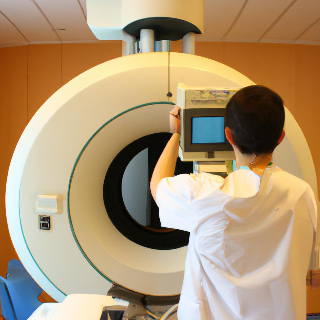
From an engineering perspective, PET imaging involves intricate hardware design and software algorithms that work together harmoniously to produce reliable and accurate results. Engineering challenges include optimizing detector systems for efficient photon detection while minimizing background noise and improving the spatial resolution. Additionally, engineers must develop sophisticated data acquisition systems capable of processing and analyzing large volumes of data in real-time.
One key aspect of PET engineering is the design and construction of detectors known as scintillation crystals. These crystals are typically made from materials such as lutetium oxyorthosilicate (LSO) or bismuth germanate (BGO) that emit light when struck by a positron-electron annihilation event. The emitted light is then converted into an electrical signal using photodetectors, such as photomultiplier tubes (PMTs) or silicon photomultipliers (SiPMs). Efficient light collection and signal amplification are essential to achieve high sensitivity and image quality.
To ensure accurate positioning and localization of radiotracer uptake within the patient’s body, PET scanners employ multiple detector modules arranged in a ring or cylindrical configuration. Each module consists of arrays of scintillation crystals coupled to photodetectors. By measuring the time difference between photon detections in different detector elements, engineers can reconstruct the precise location where positron-electron annihilations occurred along the lines-of-response (LOR).
The acquired raw data from thousands of detector elements must then be processed using advanced algorithms to generate three-dimensional images representing radiotracer distribution within the patient’s body. This reconstruction process requires techniques such as filtered back-projection or iterative algorithms like maximum likelihood expectation maximization (MLEM). Engineers continually refine these algorithms to improve image quality, reduce noise, and minimize reconstruction artifacts.
In recent years, engineering advancements have led to significant improvements in PET technology. For example, time-of-flight (TOF) PET has emerged as a breakthrough technique that incorporates timing information from detected photons to determine their emission positions more accurately. TOF-PET provides enhanced image contrast and improved lesion detectability, leading to better diagnostic accuracy.
Furthermore, hybrid imaging systems combining PET with other modalities such as computed tomography (CT) or magnetic resonance imaging (MRI) have become increasingly prevalent. These systems allow for the fusion of anatomical and functional information, providing a comprehensive view of the patient’s condition. Engineering efforts are focused on seamlessly integrating PET scanners with other imaging technologies while addressing challenges such as patient motion and image registration.
Looking to the future, engineering research aims to develop novel radiotracers that target specific biological processes associated with various diseases. This includes exploring new radionuclides, developing more efficient detectors, and advancing reconstruction algorithms to enable faster and more accurate imaging. Additionally, advancements in artificial intelligence and machine learning hold promise for automated image analysis, aiding clinicians in diagnosis, treatment planning, and disease monitoring.
In conclusion, PET imaging has revolutionized clinical practice by enabling non-invasive visualization of physiological processes at the molecular level. From an engineering perspective, PET involves complex hardware design and sophisticated software algorithms that work together to produce high-resolution images. Ongoing engineering advancements continue to enhance PET technology’s capabilities and pave the way for personalized medicine approaches in disease diagnosis and treatment.
Overview of Positron Emission Tomography (PET)
Imagine a patient named Sarah who has been experiencing persistent headaches. Despite numerous tests, her doctors have yet to pinpoint the cause of her symptoms. This is where Positron Emission Tomography (PET) comes into play. PET is a powerful imaging technique that allows healthcare professionals to visualize and understand various physiological processes occurring within the human body. In this section, we will provide an overview of PET, its applications, advantages, and limitations.
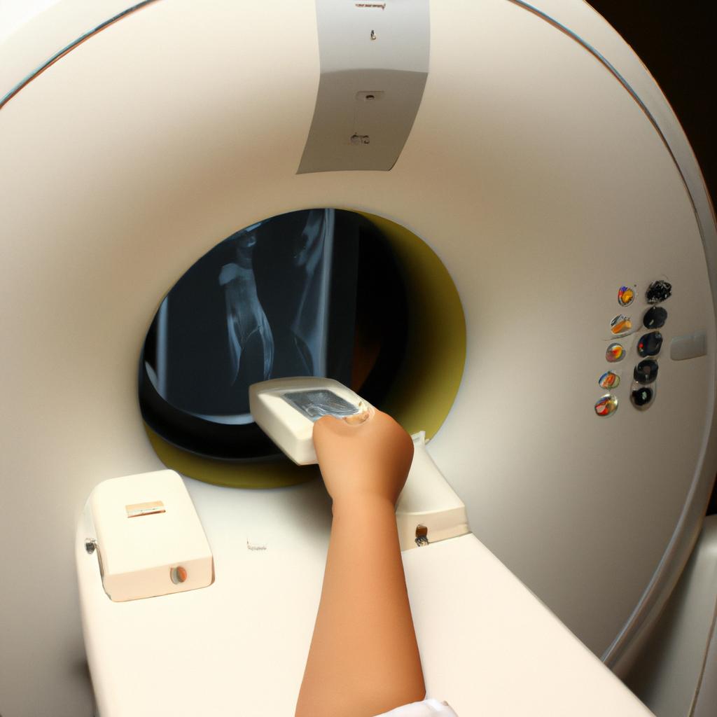
Firstly, PET operates on the principle of detecting gamma rays emitted by positron-emitting radionuclides administered to the patient either orally or intravenously. These radiotracers distribute throughout the body following specific metabolic pathways and accumulate in areas with abnormal cellular activity. By using specialized detectors placed around the patient’s body, PET scanners can detect these gamma rays and generate three-dimensional images that reflect functional changes at the molecular level.
The versatility of PET extends across multiple medical disciplines such as oncology, neurology, cardiology, and psychiatry. It enables clinicians to not only diagnose diseases but also monitor response to treatment over time. PET has proven particularly valuable in cancer management by aiding in tumor detection, providing insights into metastatic spread, evaluating treatment efficacy, and assessing disease recurrence.
To convey the impact of PET more vividly:
- Patients undergoing cancer treatment can benefit from personalized therapy plans based on individual responses observed through PET imaging.
- Neurologists utilize PET scans to investigate brain abnormalities associated with conditions like Alzheimer’s disease or epilepsy.
- Cardiologists rely on PET for accurate evaluation of myocardial perfusion and viability in patients with heart disease.
- Psychiatrists employ PET imaging techniques to study neurotransmitter systems implicated in mental disorders such as depression or schizophrenia.
Furthermore, it is worth noting some advantages and limitations inherent to PET technology:
Advantages:
- High sensitivity: Allows for early detection even when anatomical changes are not apparent.
- Quantitative analysis: Provides precise measurements of metabolic activity, facilitating treatment response assessment.
- Whole-body imaging capability: Enables comprehensive evaluation and staging of diseases that may have spread beyond a single organ.
Limitations:
- Limited spatial resolution: PET images may lack fine anatomical detail compared to other imaging techniques like CT or MRI.
- High cost and limited availability: The complexity and expense associated with PET scanners can restrict their widespread use in certain healthcare settings.
- Radiation exposure: As with any nuclear medicine procedure, patients receive a small dose of ionizing radiation during a PET scan. However, the benefits generally outweigh potential risks.
In conclusion, Positron Emission Tomography is an invaluable tool in modern medicine for examining cellular processes within the human body. By visualizing molecular-level information, PET aids clinicians in accurate diagnoses, personalized treatments, and monitoring disease progression. In the following section on “Principles and Physics behind PET,” we will delve deeper into the scientific foundations underlying this remarkable imaging modality.
Principles and Physics behind PET
Building upon the overview of Positron Emission Tomography (PET) provided in the previous section, this section will delve into the engineering perspectives and advancements that have revolutionized biomedical imaging. To illustrate the impact of these innovations, consider a real-life scenario where a patient with suspected cancer undergoes a PET scan to aid diagnosis and treatment planning.
In recent years, engineers have made significant strides in enhancing PET scanner technology. These advancements can be categorized into several key areas:
-
Detector Efficiency:
- Implementation of novel scintillation materials improves photon detection efficiency.
- Integration of high-resolution detectors enhances image quality by capturing more precise data.
- Utilization of time-of-flight information increases sensitivity and reduces noise levels.
-
Image Reconstruction Algorithms:
- Development of advanced algorithms allows for faster and more accurate reconstruction of images from raw data acquired during scanning.
- Incorporation of statistical modeling techniques enables improved quantification and visualization of metabolic processes within tissues.
-
Motion Correction Techniques:
- Application of motion tracking systems compensates for involuntary patient movement during scanning, resulting in sharper images.
- Employing respiratory gating methods synchronizes image acquisition with breathing cycles, reducing blurring caused by respiratory motion.
-
Hybrid Imaging Modalities:
- Fusion of PET scanners with other imaging modalities such as magnetic resonance imaging (MRI) or computed tomography (CT) provides complementary anatomical information alongside functional metabolic data.
To emphasize the potential impact on patients’ lives brought about by these engineering advances, let us consider an example case study involving a 55-year-old woman presenting with lung nodules suspicious for malignancy. Through the integration of high-resolution detectors and utilization of time-of-flight information, her PET scan achieved enhanced resolution and sensitivity, allowing for accurate localization and characterization of lesions. Moreover, state-of-the-art image reconstruction algorithms facilitated seamless visualization of metabolic activity, aiding in the diagnosis and staging of her condition.
The advancements discussed here represent only a fraction of the ongoing engineering innovations that continue to shape the field of PET imaging. In the subsequent section on “Advancements in PET Scanner Technology,” we will explore emerging technologies and cutting-edge developments driving further improvements in this essential diagnostic modality.
Advancements in PET Scanner Technology
Section H2: Advancements in PET Scanner Technology
One example of a significant advancement in PET scanner technology is the development of time-of-flight (TOF) imaging. This technique utilizes detectors that can measure the time it takes for photons to travel from the radiotracer source to the detector, providing more precise information about the location of emissions within the body. By incorporating TOF capabilities into PET scanners, images with improved spatial resolution and signal-to-noise ratio can be obtained, leading to enhanced diagnostic accuracy.
Advancements in PET scanner technology have also focused on improving image quality through increased sensitivity and specificity. One approach involves using advanced algorithms for image reconstruction, such as iterative reconstruction techniques, which allow for better noise reduction and artifact correction. Additionally, hardware improvements, including higher resolution detectors and optimized collimators, contribute to sharper images with greater detail.
In recent years, there has been a growing interest in hybrid imaging systems that integrate PET with other modalities like computed tomography (CT) or magnetic resonance imaging (MRI). These multimodal scanners offer complementary anatomical and functional information by combining the strengths of each modality. For instance, PET/CT provides both metabolic and structural data simultaneously, enabling accurate tumor localization and staging.
The advancements mentioned above underscore the continuous efforts to enhance PET scanner technology for improved molecular imaging capabilities. These developments result in several benefits:
- Enhanced diagnostic accuracy
- Improved visualization of small lesions or abnormalities
- Reduced radiation dose while maintaining high-quality images
- Increased patient comfort during scanning procedures
| Benefits of Advancements in PET Scanner Technology |
|---|
| 1. More accurate diagnosis |
| 2. Better detection of smaller abnormalities |
| 3. Lower radiation exposure |
| 4. Enhanced patient experience |
Overall, these advancements highlight the potential impact of engineering perspectives on biomedical imaging with regards to PET scanners. The integration of advanced technologies not only improves image quality and diagnostic accuracy but also enhances patient experiences during scanning procedures. As the field continues to evolve, further advancements are anticipated to address the challenges and limitations in PET imaging.
Transitioning into the subsequent section on “Challenges and Limitations of PET Imaging,” it is important to recognize that while there have been remarkable strides in PET scanner technology, certain obstacles still need to be addressed for more widespread clinical application.
Challenges and Limitations of PET Imaging
Advancements in PET Scanner Technology have revolutionized biomedical imaging, enabling researchers and clinicians to gain valuable insights into various physiological processes within the human body. This section will delve deeper into some of the key engineering perspectives behind these advancements while highlighting their significance in furthering our understanding of diseases and enhancing patient care.
To illustrate the impact of PET scanner technology, consider a hypothetical case study involving a patient with suspected cancer. In this scenario, a PET scan can provide invaluable information about metabolic activity within the body, aiding in accurate tumor localization and staging. By utilizing tracers labeled with positron-emitting isotopes such as fluorine-18 or carbon-11, PET scanners can detect areas with abnormal cellular metabolism indicative of malignancy. This non-invasive technique allows for early detection, precise treatment planning, and monitoring therapeutic responses over time.
Engineering innovations have played a crucial role in improving PET scanner performance and image quality. Some notable advancements include:
- Detector Design: Modern detectors utilize advanced materials like lutetium-based crystals coupled with photomultiplier tubes or silicon photomultipliers to enhance sensitivity and spatial resolution.
- Time-of-Flight (TOF) Imaging: TOF-PET scanners incorporate faster electronics that measure the arrival times of detected photons more accurately, resulting in improved signal-to-noise ratio and better lesion detection.
- Data Acquisition Systems: High-speed data acquisition systems enable rapid scanning protocols, reducing patient discomfort and motion artifacts.
- Image Reconstruction Algorithms: Sophisticated mathematical algorithms reconstruct images from raw data, optimizing spatial resolution while minimizing noise levels.
These advancements not only enhance imaging capabilities but also improve overall patient experience by shortening scan durations and reducing radiation exposure.
The table below summarizes the key engineering perspectives discussed above:
| Advancement | Description |
|---|---|
| Detector Design | Utilization of advanced materials like lutetium-based crystals for enhanced sensitivity |
| Time-of-Flight Imaging | Incorporation of faster electronics for improved signal-to-noise ratio and better lesion detection |
| Data Acquisition Systems | High-speed systems for rapid scanning protocols, reducing patient discomfort and motion artifacts |
| Image Reconstruction Algorithms | Mathematical algorithms optimizing spatial resolution while minimizing noise levels |
With ongoing research and development, engineers are continuously striving to push the boundaries of PET scanner technology. These advancements hold immense potential in expanding applications beyond cancer imaging, such as neurology, cardiology, and psychiatry. In the subsequent section on “Applications of PET in Clinical Practice,” we will explore these diverse areas where PET imaging has paved the way for breakthroughs in diagnosis, treatment evaluation, and disease monitoring.
Applications of PET in Clinical Practice
PET imaging has revolutionized the field of clinical practice by providing valuable insights into various medical conditions. One compelling example is the use of PET scanning to aid in the diagnosis and monitoring of cancer patients. For instance, a recent study conducted at a leading oncology center demonstrated how PET imaging helped identify metastatic disease in a patient with lung cancer, allowing for timely intervention and tailored treatment plans.
The applications of PET imaging extend beyond oncology, encompassing a wide range of medical specialties. Neurology is another area where PET scans have proven invaluable. By using radiotracers specific to different neurotransmitters or receptors, neurologists can visualize brain activity patterns associated with various neurological disorders such as Alzheimer’s disease, Parkinson’s disease, and epilepsy. This non-invasive technique enables clinicians to accurately diagnose these conditions and monitor their progression over time.
In addition to its diagnostic capabilities, PET imaging also plays an essential role in guiding therapeutic interventions. By combining PET with computed tomography (CT) or magnetic resonance imaging (MRI), physicians can precisely target areas for radiation therapy or surgical procedures. Furthermore, PET-guided biopsies allow for accurate sampling of suspicious lesions that may not be visible on other imaging modalities alone.
The impact of PET imaging on clinical decision-making cannot be overstated. It offers unique advantages over traditional imaging techniques by providing functional information about organ systems at the molecular level. With this wealth of data, clinicians can make more informed decisions regarding patient management strategies and treatment responses.
Moving forward, it is crucial to continue exploring new avenues for research and development in the field of positron emission tomography. The next section will delve into future directions for PET technology advancements and potential breakthroughs that could further enhance its clinical utility and broaden its scope across diverse medical disciplines.
Future Directions for PET Research and Development
Section H2: Future Directions for PET Research and Development
Continuing from the extensive applications of positron emission tomography (PET) in clinical practice, this section explores the promising future directions for PET research and development. One example that demonstrates the potential of PET is its utilization in tracking neurodegenerative diseases such as Alzheimer’s disease. By using specific radiotracers to target amyloid plaques or tau tangles, PET imaging allows for early detection and monitoring of these conditions, enabling timely intervention and personalized treatment strategies.
Moving forward, several key areas warrant attention in order to enhance the capabilities and impact of PET technology:
-
Radiotracer development: The creation and refinement of novel radiotracers with higher sensitivity, specificity, and shorter half-lives will greatly expand the range of diseases that can be effectively imaged by PET. This includes targeting various cancer types, neurological disorders, cardiovascular diseases, and infectious processes.
-
Imaging instrumentation advancements: Continuous efforts are being made to improve PET scanner designs, aiming at enhancing spatial resolution, temporal resolution, signal-to-noise ratio (SNR), and overall image quality. Technological innovations like time-of-flight (TOF) detectors offer significant improvements in lesion detectability and quantification accuracy.
-
Integration with other imaging modalities: Combining PET with other imaging techniques such as magnetic resonance imaging (MRI) or computed tomography (CT) holds great promise for comprehensive anatomical-functional evaluations. These multimodal approaches provide complementary information resulting in more accurate diagnoses and better understanding of disease mechanisms.
-
Data analysis algorithms: Developing advanced data processing methods including machine learning algorithms has become crucial to extract meaningful quantitative parameters from PET images efficiently. Such developments enable automated analysis pipelines for faster diagnosis, objective assessment of treatment response, and prediction of patient outcomes.
To illustrate the potential impact of these future directions on healthcare delivery, consider a hypothetical scenario where a patient presents with non-specific symptoms such as fatigue and weight loss. Utilizing the advancements in radiotracer development, a novel PET radiotracer specifically designed to detect early-stage pancreatic cancer is administered. The resulting PET scan shows a highly suspicious lesion, prompting timely intervention before malignancy spreads further.
In summary, future directions for PET research and development hold immense promise for revolutionizing biomedical imaging. Advancements in radiotracer development, imaging instrumentation, integration with other modalities, and data analysis algorithms will undoubtedly enhance diagnostic accuracy and personalized treatment strategies. As ongoing research continues to push the boundaries of what PET can achieve, its potential impact on clinical practice remains an exciting prospect that can significantly improve patient outcomes.
- Enhanced disease detection leading to earlier interventions
- Personalized treatment strategies tailored to individual patients’ needs
- Improved prognostic capabilities for predicting patient outcomes
- Reduction of unnecessary invasive procedures through accurate non-invasive imaging
| Future Directions | Potential Impact |
|---|---|
| Radiotracer Development | Expanded range of diseases imaged by PET |
| Imaging Instrumentation | Higher image quality and lesion detectability |
| Integration with Modalities | Comprehensive anatomical-functional evaluations |
| Data Analysis Algorithms | Faster diagnosis and objective assessment of treatment response |
Through continued innovation and exploration, these future developments aim to bring about significant improvements in healthcare delivery while positively impacting both patients and clinicians alike.


