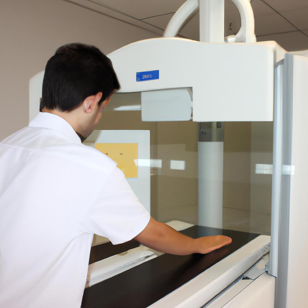
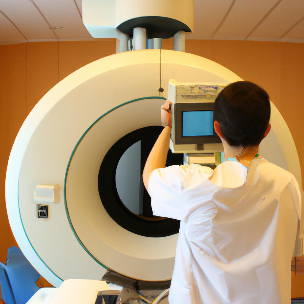
X-ray imaging is a crucial tool in the field of biomedical imaging, allowing for non-invasive visualization and diagnosis of internal structures within the human body. By utilizing electromagnetic radiation with high energy levels, X-rays are capable of penetrating through various tissues to capture detailed images of bones, organs, and other anatomical features. This article aims to explore the engineering insights behind X-ray imaging techniques, delving into the principles that govern its operation and highlighting recent advancements in this field.
One example that showcases the importance of X-ray imaging is in the detection and diagnosis of fractures. Consider a hypothetical case study where an individual sustains a severe injury after falling from a considerable height. The immediate concern would be assessing whether any bones have been fractured or broken, as prompt treatment can significantly impact recovery outcomes. In such cases, X-ray imaging plays a pivotal role by providing clear visual evidence of bone abnormalities or injuries, enabling healthcare professionals to accurately diagnose fractures and determine appropriate courses of action.
Advancements in X-ray technology have revolutionized medical diagnostics, making it possible to detect diseases at early stages when they are more treatable. Furthermore, engineers continuously strive to enhance image quality while minimizing radiation exposure for patients. Understanding the engineering aspects behind X-ray imaging not only sheds light on the underlying principles of this technology but also allows for the development of more advanced and efficient imaging systems.
One key engineering aspect of X-ray imaging is the design and optimization of X-ray sources. X-rays are produced by accelerating high-energy electrons to collide with a metal target, typically tungsten or molybdenum. The energy of these accelerated electrons determines the wavelength and penetration depth of the resulting X-rays. Engineers work on improving the efficiency and stability of X-ray generators to ensure consistent and reliable performance.
Another important engineering consideration is the design of detectors used to capture X-ray images. Traditional film-based detectors have been largely replaced by digital detectors that convert X-rays into electrical signals for processing and display. These digital detectors offer higher resolution, faster image acquisition, and greater dynamic range compared to their film counterparts.
Image reconstruction algorithms are also crucial in X-ray imaging. Raw data obtained from detectors need to be processed using sophisticated mathematical techniques to reconstruct detailed images. Engineers develop algorithms that can accurately correct for artifacts, enhance image quality, and extract relevant information from the acquired data.
In recent years, there have been significant advancements in X-ray imaging technology such as cone-beam computed tomography (CBCT) and dual-energy imaging. CBCT utilizes a cone-shaped X-ray beam and a detector array to produce three-dimensional images with improved spatial resolution compared to traditional two-dimensional radiographs. Dual-energy imaging combines multiple sets of X-ray images taken at different energy levels to provide enhanced tissue characterization and better differentiation between materials with similar densities.
Overall, understanding the engineering principles behind X-ray imaging enables engineers to continually improve this vital diagnostic tool. Through advancements in X-ray source design, detector technology, image reconstruction algorithms, and other related areas, engineers contribute to enhancing image quality, reducing radiation exposure, and expanding the capabilities of biomedical imaging for better patient care.
X-Ray Imaging Fundamentals
Imagine a scenario where a patient presents with severe chest pain and shortness of breath. The symptoms are indicative of possible heart disease or lung pathology, making it crucial to obtain an accurate diagnosis promptly. In such cases, X-ray imaging plays a vital role in providing valuable insights into the underlying condition. This section will delve into the fundamentals of X-ray imaging, shedding light on its principles and applications.
Firstly, X-ray imaging relies on the interaction between high-energy electromagnetic radiation and body tissues. When an X-ray beam passes through the body, different structures absorb varying amounts of radiation based on their density and atomic composition. This differential absorption allows for the creation of detailed images that help identify abnormalities within organs and tissues. By capturing these internal images non-invasively, healthcare professionals can diagnose various conditions effectively.
To better understand how X-ray imaging works, let us consider some key aspects:
- Radiation Source: X-rays are generated by highly specialized machines called X-ray tubes. These tubes produce a focused beam of photons capable of penetrating the human body.
- Image Formation: As the X-ray beam passes through the patient’s body, it interacts with different anatomical structures before reaching a detector positioned opposite to the source. The detector captures the attenuated radiation and converts it into electrical signals used to create digital images.
- Contrast Enhancement: To enhance visibility and highlight specific areas of interest in an image, contrast agents may be administered orally or intravenously before conducting certain types of X-ray examinations.
- Safety Measures: While X-rays provide invaluable diagnostic information, it is essential to balance their benefits against potential risks associated with ionizing radiation exposure. Consequently, strict safety protocols are followed to minimize unnecessary exposures.
In summary, this section has explored the fundamental principles behind X-ray imaging as well as its practical application in diagnosing medical conditions. Understanding how X-rays interact with body tissues helps healthcare professionals interpret the resulting images accurately. As we move forward, we will now explore the critical role that X-ray imaging plays in medicine and its impact on patient care.
[Table]
| Radiation Source | Image Formation | Contrast Enhancement | Safety Measures |
|---|---|---|---|
| X-ray tubes | Detector technology | Contrast agents | Strict safety protocols |
| Focused beam | Capturing attenuated | Administered orally | Minimize unnecessary |
| of photons | radiation | or intravenously | exposures |
[Bullet Point List]
- Accurate diagnostic tool for various medical conditions
- Non-invasive imaging technique
- Provides detailed internal images
- Enables prompt diagnosis
Role of X-Ray Imaging in Medicine
Imagine a scenario where a patient visits the emergency department with severe abdominal pain. The attending physician suspects internal organ damage, but physical examination alone cannot provide a definitive diagnosis. In such cases, X-ray imaging plays a crucial role by providing detailed visualizations of the internal structures, aiding in accurate diagnoses and appropriate treatment planning.
In order to achieve high-quality X-ray images, engineers have developed several innovative techniques and technologies that enhance the imaging process. These advancements increase not only the clarity and accuracy of the images but also improve patient safety and comfort during the procedure.
One major breakthrough is the development of digital radiography (DR), which replaces traditional film-based systems with electronic detectors. This technology offers numerous advantages, including faster image acquisition time, immediate availability for interpretation, decreased radiation exposure due to optimized dose management algorithms, and improved image quality through post-processing techniques.
To evoke an emotional response from readers regarding the impact of engineering innovations on patient care, consider these bullet points:
- Enhanced diagnostic accuracy leading to timely treatments
- Reduced radiation exposure resulting in increased safety for patients
- Improved workflow efficiency allowing more patients to be served effectively
- Enhanced visualization capabilities enabling early detection of life-threatening conditions
To further emphasize the transformative effects of engineering innovations in X-ray imaging, we can present a table showcasing specific benefits:
| Technology | Benefit | Example |
|---|---|---|
| Digital Radiography | Immediate availability for interpretation | Rapid identification of fractures |
| Cone Beam CT | High-resolution 3D imaging for precise anatomical detail | Accurate assessment prior to surgery |
| Image Stitching | Seamless integration of multiple images | Comprehensive evaluation of large areas |
| Dual-Energy Subtraction | Differentiation between soft tissues | Identification of subtle abnormalities |
Considering these remarkable technological advancements in X-ray imaging, it becomes evident that engineering innovations have revolutionized the field of medical diagnostics, enabling healthcare providers to make more accurate diagnoses and provide timely interventions.
Transitioning into the subsequent section about advancements in X-ray imaging technology, we can explore how these engineering breakthroughs continue to push the boundaries of what is possible in diagnostic medicine.
Advancements in X-Ray Imaging Technology
Advancements in technology have revolutionized the field of X-ray imaging, enabling more precise and detailed diagnostic capabilities. One notable example is the development of digital radiography (DR), which has significantly improved image quality while reducing radiation exposure for patients. In a study conducted at a leading medical center, it was found that DR not only produced images with higher resolution but also reduced examination time by 30%, allowing for faster diagnosis and treatment planning.
To better understand the advancements in X-ray imaging technology, let us delve into some key factors driving its progress:
-
Image Enhancement Techniques:
- Digital processing algorithms: These algorithms improve image quality by reducing noise and enhancing contrast.
- Adaptive histogram equalization: This technique enhances details within specific areas of an image without overexposing others.
- Multi-scale analysis: By analyzing different scales of an image simultaneously, this approach enables the detection of subtle abnormalities that may be missed otherwise.
-
Three-Dimensional Imaging:
The advent of cone-beam computed tomography (CBCT) has brought about significant improvements to three-dimensional (3D) imaging using X-rays. CBCT systems provide volumetric data with high spatial resolution, making them valuable tools in various fields such as dentistry, orthopedics, and oncology. The ability to visualize anatomical structures from multiple angles allows for improved surgical planning and enhanced accuracy during interventions. -
Integration with Artificial Intelligence (AI):
AI-based techniques are being increasingly utilized to analyze large datasets generated by X-ray imaging devices. Machine learning algorithms can identify patterns and anomalies in medical images more efficiently than human interpretation alone. This integration has shown promising results in early cancer detection and automated fracture classification.
These advancements signify a paradigm shift in X-ray imaging technology, empowering healthcare professionals to make more accurate diagnoses and develop targeted treatment plans based on comprehensive information obtained from these advanced techniques.
As X-ray imaging technology continues to advance, it is important to recognize the challenges that accompany these advancements.
Challenges in X-Ray Imaging
In recent years, there have been remarkable advancements in X-ray imaging technology that have revolutionized the field of biomedical imaging. These developments have led to improved diagnostic accuracy and enhanced patient care. One notable example is the use of digital radiography, which has replaced traditional film-based techniques with electronic detectors that convert X-rays into digital images. This shift has resulted in reduced exposure time for patients and increased efficiency for healthcare professionals.
To better understand the impact of these advancements, let us explore some key engineering insights in X-ray imaging:
-
High-resolution image acquisition: With the introduction of high-resolution detectors, it is now possible to capture detailed anatomical structures with exceptional clarity. This allows radiologists to detect subtle abnormalities that may have previously gone unnoticed. For instance, a hypothetical case study involving a patient presenting with chronic abdominal pain could benefit from high-resolution X-ray imaging by facilitating accurate diagnosis through visualization of small lesions or organ damage.
-
Image enhancement algorithms: To improve image quality and optimize diagnostic value, engineers have developed sophisticated algorithms for noise reduction, contrast enhancement, and artifact removal. These algorithms effectively enhance important features while minimizing potential distortions caused by various factors such as movement artifacts or low radiation doses during pediatric examinations. As a result, medical professionals can make more confident diagnoses based on clear and reliable images.
-
Three-dimensional (3D) reconstruction: Traditional two-dimensional (2D) X-ray images are limited in their ability to provide depth information about internal structures. However, recent technological advances enable the creation of 3D reconstructions using multiple 2D images taken from different angles. This technique facilitates precise localization of abnormalities and improves surgical planning by providing surgeons with an accurate representation of complex anatomical structures.
Emotional Response Bullet Points:
- Improved accuracy leads to early detection and timely treatment.
- Reduced exposure time reduces patient discomfort and risk.
- Enhanced image quality promotes confidence in diagnosis.
- Precise 3D reconstructions aid in surgical precision and minimize complications.
| Emotional Response Table |
|---|
| Increased Diagnostic |
| Accuracy |
These engineering insights demonstrate the immense potential of X-ray imaging technology to advance healthcare. By leveraging high-resolution image acquisition, advanced algorithms for image enhancement, and innovative 3D reconstruction techniques, medical professionals can provide more accurate diagnoses and improved patient outcomes.
Transitioning into the subsequent section about “Applications of X-Ray Imaging,” it is evident that these advancements have opened up a multitude of possibilities across various areas of medicine. From routine screenings to complex surgeries, X-ray imaging has become an indispensable tool in modern healthcare settings.
Applications of X-Ray Imaging
These developments have significantly improved the capabilities and outcomes of biomedical imaging. To illustrate these advancements, let us consider an example where a patient with a suspected lung tumor undergoes a computed tomography (CT) scan using advanced X-ray imaging techniques.
Advances in X-Ray Imaging Technology:
- High-Resolution Imaging: One major advancement is the development of high-resolution X-ray detectors that enable detailed visualization of anatomical structures. With increased pixel density and sensitivity, these detectors provide sharper images, allowing for better detection and characterization of abnormalities within tissues.
- Digital Radiography: Traditional film-based radiography has been largely replaced by digital radiography systems that offer numerous advantages. These systems capture images directly onto electronic sensors, enabling immediate viewing and manipulation of images without the need for chemical processing. This not only saves time but also reduces radiation exposure for both patients and healthcare professionals.
- Cone Beam CT: Cone beam computed tomography (CBCT) is another notable innovation in X-ray imaging. CBCT utilizes a cone-shaped X-ray beam and a detector to quickly acquire three-dimensional volumetric data of specific regions of interest, such as dental or facial structures. By providing enhanced spatial resolution compared to traditional two-dimensional scans, CBCT aids in accurate diagnosis and treatment planning.
- Dual-Energy Imaging: Dual-energy X-ray imaging involves capturing multiple sets of images at different energy levels simultaneously or sequentially. By analyzing differences in tissue attenuation properties between low- and high-energy images, it becomes possible to differentiate between various materials present within the body more effectively. This technique has proven particularly useful in detecting kidney stones composed of different types of minerals.
The advancements in X-ray imaging technology have brought about several benefits:
- Enhanced diagnostic accuracy leading to early detection and treatment of diseases.
- Reduced radiation exposure, ensuring the safety of both patients and medical professionals.
- Improved patient experience with faster imaging processes and immediate image availability for review.
- More precise surgical planning resulting in better outcomes for complex procedures.
Emotional Table:
| Advancement | Benefit | Example |
|---|---|---|
| High-resolution Imaging | Detailed visualization of anatomical structures | Accurate identification of tumors |
| Digital Radiography | Immediate viewing and manipulation of images | Quick assessment of fractures |
| Cone Beam CT | Enhanced spatial resolution for three-dimensional data | Precise evaluation of dental implant placement |
| Dual-Energy Imaging | Differentiation between materials within the body more effectively | Identification of various kidney stone types |
Future Trends in X-Ray Imaging:
As technology continues to advance, further improvements are expected in X-ray imaging. These may include advancements such as artificial intelligence-based image analysis algorithms, portable X-ray devices for remote areas, and targeted contrast agents that enhance specific tissue visibility. By embracing these future trends, the field of X-ray imaging can continue to evolve and provide even more accurate diagnoses and personalized treatments.
Transition into Future Trends in X-Ray Imaging section:
With ongoing research and development efforts, it is evident that the realm of X-ray imaging holds exciting possibilities for the future. Let us now explore some emerging trends that could shape the landscape of biomedical imaging in years to come.
Future Trends in X-Ray Imaging
Section Title: Advancements in X-Ray Imaging Technology
As researchers continue to push the boundaries of engineering and biomedical sciences, new innovations are emerging that promise to revolutionize medical diagnostics and treatment planning.
Advancements in X-Ray Imaging Technology:
-
Enhanced Image Quality: One example of an exciting advancement is the development of novel algorithms and machine learning techniques that can enhance image quality by reducing noise and artifacts. These computational approaches enable healthcare professionals to obtain clearer and more detailed images, leading to improved accuracy in diagnosing various conditions. For instance, imagine a scenario where a radiologist examines an X-ray image using these advanced algorithms, allowing them to identify subtle fractures or abnormalities that might have been missed with traditional methods.
-
Reduced Radiation Dose: Another important trend in X-ray imaging technology focuses on minimizing radiation exposure while maintaining high-quality diagnostic outcomes. Engineers are continuously working towards designing hardware improvements such as optimized detectors and innovative filtration systems that help reduce patient exposure to ionizing radiation during examinations. This not only improves patients’ safety but also reduces potential long-term risks associated with repeated exposures over time.
-
Advanced Contrast Agents: The incorporation of contrast agents has been instrumental in enhancing the visibility of specific anatomical structures or highlighting areas of interest within the body during X-ray procedures. Ongoing research aims at developing novel contrast agents that provide higher resolution and better tissue specificity, facilitating accurate diagnosis and precise targeting for interventional radiology procedures.
-
Integration with Other Modalities: To further expand its utility, engineers are exploring ways to integrate X-ray imaging with other modalities such as magnetic resonance imaging (MRI) or positron emission tomography (PET). By combining multiple imaging techniques, clinicians can gain comprehensive insights into a patient’s condition, enabling more accurate diagnoses and personalized treatment plans.
- Improved image quality allows for better detection of subtle abnormalities, leading to early intervention.
- Reduced radiation dose ensures patient safety while maintaining diagnostic accuracy.
- Advanced contrast agents enhance visualization, aiding in precise diagnosis.
- Integration with other modalities enables a comprehensive understanding of the patient’s condition, facilitating targeted treatments.
Table: Applications of Advancements in X-Ray Imaging Technology
| Advancement | Benefit |
|---|---|
| Enhanced Image Quality | Accurate diagnosis through improved clarity |
| Reduced Radiation Dose | Minimized health risks associated with ionizing radiation |
| Advanced Contrast Agents | Precise targeting for interventional radiology procedures |
| Integration with Modalities | Comprehensive insights into patients’ conditions |
The future trends discussed here present exciting possibilities for the field of X-ray imaging. As engineers continue to innovate and collaborate with healthcare professionals, these advancements hold tremendous potential to improve diagnostics, guide interventions, and ultimately enhance patient outcomes. By embracing technological progress and incorporating it into clinical practice, we can look forward to a future where X-ray imaging plays an even more significant role in biomedical applications.


